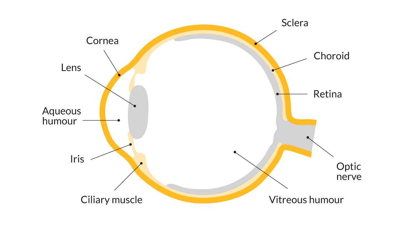
Retina Diagram Eye. It consists of photoreceptor cells (e.g. When an ophthalmologist uses an ophthalmoscope to look into your eye he sees the following view of the retina (fig. the retina is the innermost layer of the eye. the retina is approximately 0.5 mm thick and lines the back of the eye. the retina constitutes the inner layer (internal tunic) of the eyeball, found anterior to the choroid and posterior to the vitreous body. It's composed of several layers, including one that contains specialized. It starts the posterior surface of the eyeball and terminates anteriorly at the ora serrata. the retina converts light that enters into your eye into electrical signals your optic nerve sends to your brain which creates the images you see. the retina is the sensory membrane that lines the inner surface of the back of the eyeball. In the center of the retina is. It’s a key part of your. The optic nerve contains the ganglion cell axons. Rods and cones) that convert light energy into a nerve impulse.
from www.sightsavers.org
It consists of photoreceptor cells (e.g. In the center of the retina is. the retina converts light that enters into your eye into electrical signals your optic nerve sends to your brain which creates the images you see. Rods and cones) that convert light energy into a nerve impulse. It’s a key part of your. The optic nerve contains the ganglion cell axons. the retina is the innermost layer of the eye. It's composed of several layers, including one that contains specialized. the retina constitutes the inner layer (internal tunic) of the eyeball, found anterior to the choroid and posterior to the vitreous body. It starts the posterior surface of the eyeball and terminates anteriorly at the ora serrata.
How do the eyes work? Parts of the eye Sightsavers
Retina Diagram Eye the retina is approximately 0.5 mm thick and lines the back of the eye. In the center of the retina is. It starts the posterior surface of the eyeball and terminates anteriorly at the ora serrata. The optic nerve contains the ganglion cell axons. It consists of photoreceptor cells (e.g. the retina is the sensory membrane that lines the inner surface of the back of the eyeball. It’s a key part of your. the retina converts light that enters into your eye into electrical signals your optic nerve sends to your brain which creates the images you see. the retina is the innermost layer of the eye. When an ophthalmologist uses an ophthalmoscope to look into your eye he sees the following view of the retina (fig. the retina constitutes the inner layer (internal tunic) of the eyeball, found anterior to the choroid and posterior to the vitreous body. Rods and cones) that convert light energy into a nerve impulse. the retina is approximately 0.5 mm thick and lines the back of the eye. It's composed of several layers, including one that contains specialized.